-
[ February 17, 2014]
Progress made in the research of super-resolution microscopy
-
Recently, Dr. Peng Xi ’s group in Department of Biomedical Engineering, College of Engineering, realized super-resolution imaging of NV centres in the same specimen utilizing stimulated emission depletion and structured illumination microscopies, in collaboration with Dr. Wen-Di Li’s group at University of Hong Kong, Prof. Huan-Cheng Chang’s group at Taiwan, and Dr. Lei Huang at Tsinghua University.
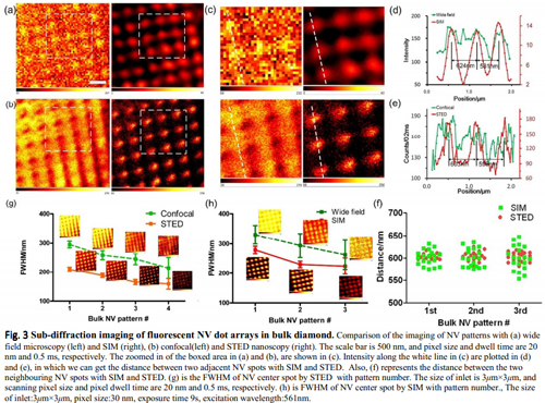
Stimulated emission depletion (STED) and structured illumination (SIM) are two commonly used techniques for super-resolution imaging. However, the performance of these two techniques has not yet been quantitatively compared side-by-side. Taking advantage of the non-photobleaching characteristic of NV centers in fluorescent nanodiamond (FND), Xi group has performed a comparative study for the resolution of these two methods with 35 nm FNDs at the single particle level, as well as with FND grown in bulk diamond material. Results show that STED provides more structural details, whereas SIM provides a larger field of view with a higher imaging speed. SIM may introduce deconvolution smooth and orientational artifacts during its post-processing. The results were published on RSC Advances entitled” Sub-diffraction imaging of nitrogen-vacancy centers in diamond by stimulated emission depletion and structured illumination” (link: http://pubs.rsc.org/En/content/articlelanding/2014/ra/c3ra47240j#!divAbstract)
Xusan Yang from Peng’s group is the first author of this paper, and Peng Xi is corresponding author. Xi’s research interests are on sub-diffraction optical nanoscopy, laser scanning confocal microscopy, multiphoton microscopy and optical coherence tomography. (http://bme.pku.edu.cn/~xipeng)